What’s the diagnosis?
A baby with a yellow-red lesion on the back
Case presentation
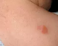
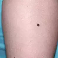
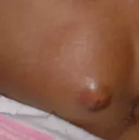
A 9-month-old girl presents with a raised lesion on her back (Figure 1). The lesion has a light yellow to orange hue and is of recent onset. She is not bothered by it and is otherwise well.
Differential diagnosis
Conditions to include in the differential diagnosis for a child of this age include the following.
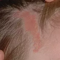
- Sebaceous naevus. This benign tumour of sebaceous gland origin occurs sporadically and is present from birth, although sometimes it is not noticed for a few weeks. Lesions are characterised by slightly raised, hairless plaques that are well defined and waxy in texture and have a yellow-orange and/or pink colour (Figure 2). They can be round, oval or linear in shape and they vary in size and number. This diagnosis should be considered for the lesion in Figure 1, but a sebaceous naevus will more commonly occur in locations of greater numbers of sebaceous glands, such as on the scalp and face. This girl’s lesion was not present at birth, so a sebaceous naevus can be excluded from the diagnosis.
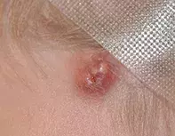
- Pilomatrixoma. This benign tumour derived from hair matrix cells can present at any time in childhood but it is relatively uncommon.1 Typically skin-coloured and solitary, a pilomatrixoma is papular or nodular and contains calcium, which makes it firm to touch (Figure 3). It occurs in the dermis or subcutaneous tissue and is covered by normal skin, most commonly on the head, neck or upper extremities. A blue discolouration is quite common. A pilomatrixoma should be considered for the lesion described above, but a yellow colour would be highly unusual and its location make this diagnosis unlikely.
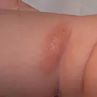
- Mastocytoma. Relatively common in infants, a mastocytoma is an asymptomatic benign collection of excess mast cells. It is characterised by red to golden-brown papules or plaques and bullae formation and blistering, particularly in areas susceptible to friction (Figure 4). Stroking or rubbing causes the lesion to swell, with increased erythema and a surrounding wheal and flare due to histamine release (Darier’s sign);2 this is occasionally accompanied by flushing or urticaria.2 A mastocytoma can be present at birth but most cases appear in the first two years in of life. They tend to involute during childhood.
- Spitz naevus. This uncommon, benign, asymptomatic melanocytic lesion usually occurs in children. It is typically brown or red in colour with a smooth surface (Figure 5), and it is usually more papular or nodular in shape than the lesion shown in Figure 1. Most Spitz naevi are less than 1 cm in diameter. They have a characteristic history of rapid growth before stabilising, and in some cases they spontaneously regress.
- Epidermoid cyst. This cyst (also known as an sebaceous cyst) has an epidermal lining and is filled with keratin (Figure 6). The lesion, which tends to be located on the head, chest or upper back, is nodular or spherical in shape and is located in the dermis or subcutaneous tissue. It very rarely affects children, making it highly unlikely in this scenario.
- Juvenile xanthogranuloma. This is the correct diagnosis. Juvenile xanthogranuloma is a benign tumour of histiocytic cells that usually occurs in infants and young children, with unknown cause.3 The lesion typically develops rapidly and is usually solitary, but some patients present with multiple lesions. The face, neck, scalp and upper parts of the body are most commonly affected. The lesion will often first appear as a red and/or yellow papule that progresses to a yellow-brown plaque or macule. It is usually firm and rubbery to touch, with small and flat papules, and there may be evidence of telangiectasia. On dermoscopy, a juvenile xanthogranuloma appears amorphous and yellow. Cutaneous lesions can be accompanied by lesions involving other organs, such as the liver, lung, spleen, testes, pericardium, gastrointestinal tract, kidneys, central nervous system and eyes. For the patient described above, the colour, texture and onset of the lesion as well as the girl’s age are strong indicators of juvenile xanthogranuloma.
Investigations
Juvenile xanthogranuloma is generally a clinical diagnosis and a biopsy is rarely performed, but the diagnosis can be confirmed on histology. Subcutaneous lesions are very difficult to diagnose clinically because they simply present with a deep nodule and biopsy is often required to make a diagnosis.
Management
Juvenile xanthogranuloma does not require specific treatment and parents should be reassured that the tumour is benign. The natural history is for the tumour to self-resolve over one to five years, and a small area of hyperpigmentation or atrophy may be left behind. It is standard practice to refer children with a cutaneous juvenile xanthogranuloma to an ophthalmologist to check for ocular lesions – these are very rare but should be excluded because they may increase intraocular pressure. Lesions in other noncutaneous locations usually do not cause any problems and will resolve spontaneously.
References
1. Schlechter R, Hartsough NA, Guttman FM. Multiple pilomatricomas (calcifying epitheliomas of Malherbe). Pediatr Dermatol 1984; 2: 23-25.
2. Soter NA. Mastocytosis and the skin. Hematol Oncol Clin North Am 2000; 14: 537-555.
3. Janssen D, Harms D. Juvenile xanthogranuloma in childhood and adolescence: a clinicopathologic study of 129 patients from the kiel pediatric tumor registry. Am J Surg Pathol 2005; 29: 21-28.
Skin lesions

